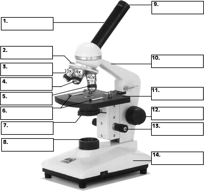
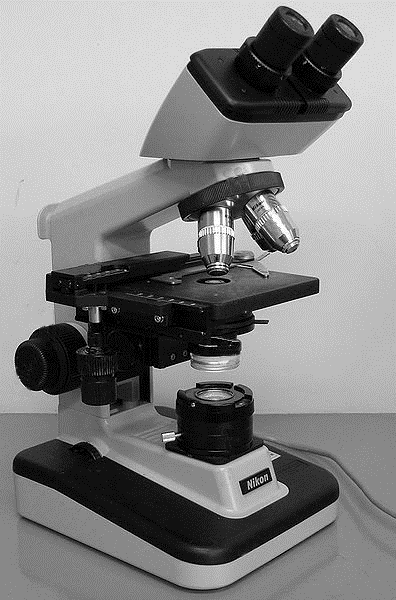
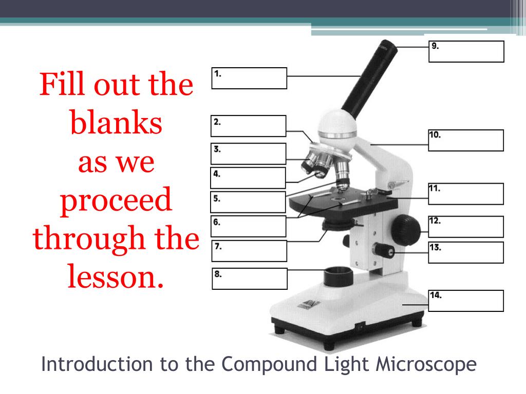
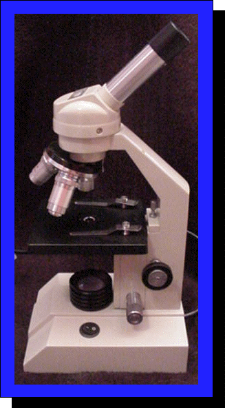
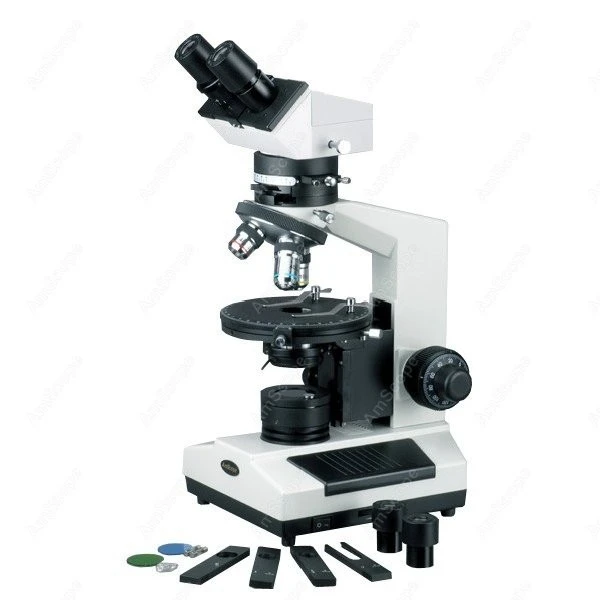
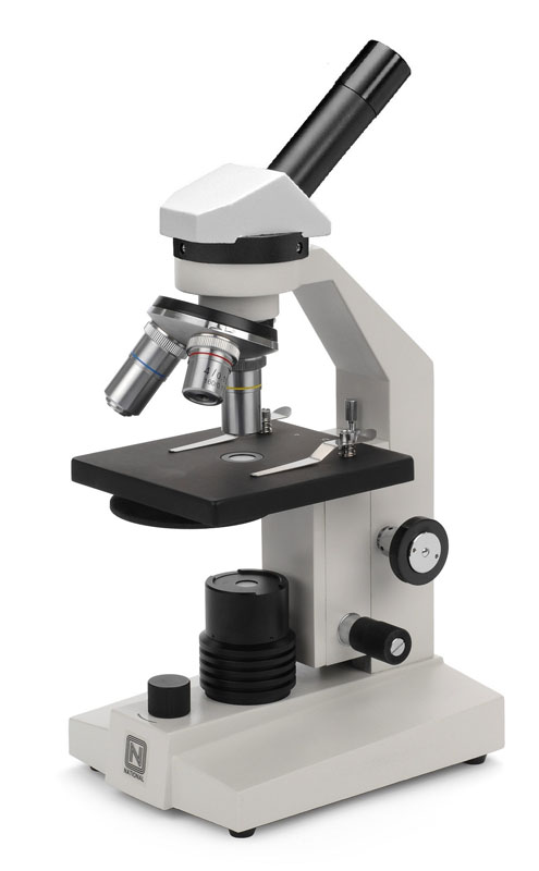
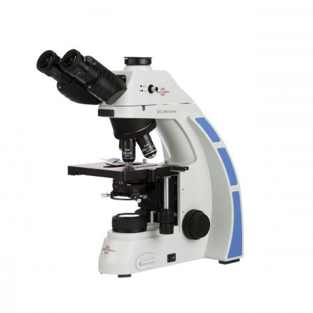
40 compound microscope unlabeled
Enzymatic Immunohistochemistry | SpringerLink The indirect method involves an initial reaction with an unlabeled primary antibody. In the next step, a second labeled antibody reacts with the primary unlabeled antibody leading to an amplified signal. The most commonly employed enzyme labels include horseradish peroxidase (HRP), alkaline phosphatase (AP) or glucose oxidase. Compound Light Microscope Diagram Worksheet - Google Groups You will label sketches to compound light microscope worksheet may want to your students to use worksheets to. On a typical student compound light microscope there are 3-4 of objective lenses. In...
A new carnivorous plant lineage (Triantha) with a unique sticky ... Drosophila melanogaster (unlabeled) 3.99 ± 0.08: 9.47 ± 0.05: 4: Drosophila melanogaster (labeled) 9.15 ± 0.39: 6: ... (Leica DMR compound microscope equipped with an EBQ 100 isolated mercury lamp; excitation, 450 to 490 nm; dichromatic mirror, DM 510 nm; emission filter, 510 nm). Fluorescence was documented with a Canon EOS Rebel T5 digital ...
Compound microscope unlabeled
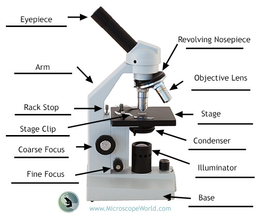
Microscope Parts | A Guide on their Location and Function A compound microscope consists of parts that assist in viewing with a naked eye, a sample holder, a magnifying lens, and a light source. Microscope Parts in detail There are almost 15 parts in a microscope. But we can divide them based on their purpose in the instrument like A) Parts that assist in viewing the object BIO 4150 HUMAN ANATOMY AND PHYSIOLOGY COURSE PROCEDURE - Cowley College ... Label the segments of an unlabeled ECG and match each event to a cardiac cycle occurrence. Given any two of three variables (cardiac output, heart rate, and stroke volume), solve for the value of the third. Given a random list of cardiac cycle events, rank the list in their logical order of occurrence. Explain the functions of the lymphatics. Caveat fluorophore: an insiders' guide to small-molecule ... - Nature The development of new microscopes, fluorescent labels and analysis techniques has pushed the frontiers of biological imaging forward, moving from fixed to live cells, from diffraction-limited to ...
Compound microscope unlabeled. how can a light microscope work - Shamika Mcgill A light microscope works by gathering a focused visible beam of light that passes through and around a specimen with the objective lens the lens closest to the specimen. Slowly lift the stage in order to get a stronger focus. Both the rheostat on. A light microscope is a microscope that uses light to view objects. An objective lens and an eyepiece. printable unlabeled brain diagram Coloring brain diagram psychology ... 14 Best Images Of Human Anatomy Labeling Worksheets - Blank Head And microscope parts worksheet label diagram compound printable science worksheets labeling quiz labeled biology light microscopes worksheeto anatomy human simple use Kid Corner - Infant And Child Studies At The University Of Maryland childstudies.umd.edu Preclinical evaluation of FAP-2286 for fibroblast ... - SpringerLink Images were acquired by using the BZ X800E fluorescence microscope (Keyence) at 40 × magnification. ... and an excess of 5 µM unlabeled competitor compound was used for blocking (bottom images) (B). Cells were further incubated for an additional 1, 3, 8, 24, and 72 h (C). For visualization of lysosomal compartments, LysoTracker DeepRed was used. Student Worksheet For Microslide - Google Groups Digital microscope BUNDLE example of a Compound light microscope and not intimidated by it, videos, stage! Conceptually, Agreement Of Adjectives Spanish Worksheet Answers Hayes School once a learning moderate for teaching pupils storage on lessons learned in the classroom. ... Rahul looks at six unlabeled slides showing different stages of the ...
Microscope Quiz: How Much You Know About Microscope Parts ... - ProProfs Projects light upwards through the diaphragm, the specimen, and the lenses. 5. Is used to regulates the amount of light on the specimen. Supports the slide being viewed. Moves the stage up and down for focusing. 6. Is used to support the microscope when carried. Moves the stage slightly to sharpen the image. Super‐Resolution Microscopy Using a Bioorthogonal‐Based Cholesterol ... To assess whether the chemical modification introduced in this probe interferes with the native behavior of the natural compound, ... identical results were obtained to those observed with the unlabeled ... Manheim, Germany) equipped with the STED white 100x NA1, 40 oil objective. The microscope was equipped with 3 depletion lines and for this ... Next-Generation Laser Scanning Multiphoton Microscopes ... - Cambridge Core The entire microscope consists of two parts that are connected through a flexible umbilical cord. The control box is on wheels and incorporates a built-in PC together with other electronic and photonic devices. ... Figure 5 shows unlabeled FFPE sections of a melanoma (A) ... It is critical to ensure the treatment compound, for example ... Deep Learning for Reconstructing Low-Quality FTIR and Raman Spectra─A ... Herein we report on a deep-learning method for the removal of instrumental noise and unwanted spectral artifacts in Fourier transform infrared (FTIR) or Raman spectra, especially in automated applications in which a large number of spectra have to be acquired within limited time. Automated batch workflows allowing only a few seconds per measurement, without the possibility of manually ...
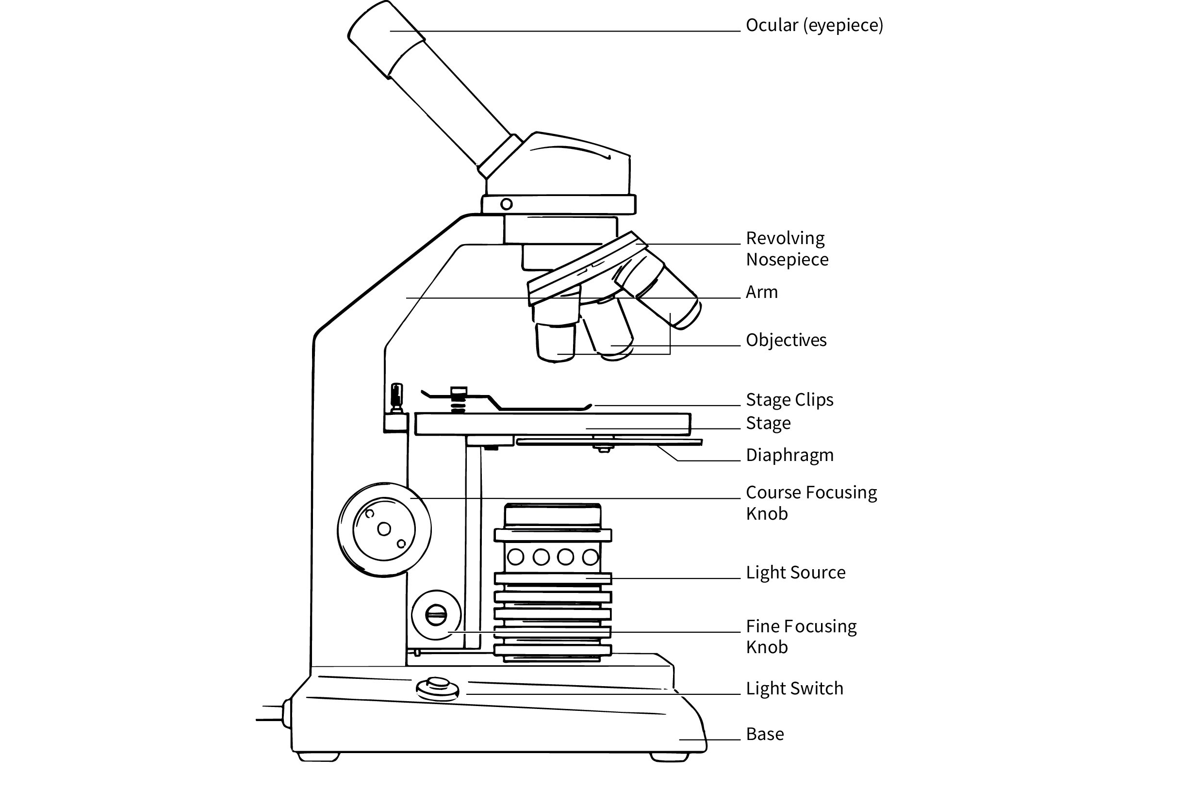
diagram shows a typical light microscope with its parts labeled ... _ocular lens (eyepiece) body tube revolving nosepiece objectives arm stage clips stage coarse adjustment knob fine adjustment knob diaphragm light source base after you learned these parts, copy and paste the following unlabeled diagram onto assignment box, and identify the parts: _ocular lens (eyepiece) body tube revolving nosepiece objectives … Chale9128 Psychology Final Exam - ProProfs Quiz 14. A man from India is experiencing fatigue, loss of appetite, weakness, anxiety, and sexual dysfunction. He is also feeling extremely guilty due to his fear of losing semen during nocturnal emissions. This culture-bound syndrome is known as : A. Dhat. Development of an intracellular quantitative assay to measure compound ... This model allows the determination of k on and k off of unlabeled test compounds in the presence of a competing pre-characterized labeled ligand. This type of approach is scalable to enable routine test compound characterisation; however, it has yet to be utilized to measure drug-target binding kinetics for intracellular targets in live cells ... Compound Microscope- Definition, Labeled Diagram, Principle, Parts, Uses A compound microscope is of great use in pathology labs so as to identify diseases. Various crime cases are detected and solved by drawing out human cells and examining them under the microscope in forensic laboratories. The presence or absence of minerals and the presence of metals can be identified using compound microscopes.
Microscope Head Unlabeled Olympus WHC10X-H /20 Japan | eBay Microscope Head Unlabeled Olympus WHC10X-H /20 Japan: Condition: For parts or not working. Ended: Jun 15, 2022. Winning bid: US $178.50 [ 9 bids] Shipping: $13.95 Standard ... Olympus Compound Microscope Binocular Microscopes, Confocal Microscope Microscopes, Microscopes, Trinocular Microscopes, Digital Microscopes; Additional site navigation.
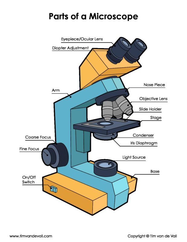
Parts of a microscope with functions and labeled diagram Microscopes are instruments that are used in science laboratories to visualize very minute objects such as cells, and microorganisms, giving a contrasting image that is magnified. Microscopes are made up of lenses for magnification, each with its own magnification powers.
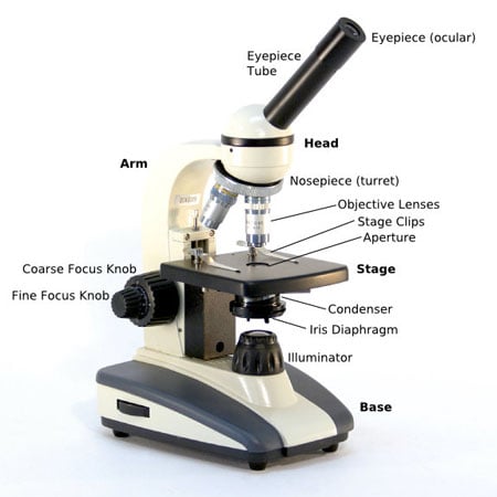
Parts of the Microscope with Labeling (also Free Printouts) Parts of the Microscope with Labeling (also Free Printouts) A microscope is one of the invaluable tools in the laboratory setting. It is used to observe things that cannot be seen by the naked eye. Table of Contents 1. Eyepiece 2. Body tube/Head 3. Turret/Nose piece 4. Objective lenses 5. Knobs (fine and coarse) 6. Stage and stage clips 7. Aperture
In vivo tracking of unlabelled mesenchymal stromal cells by ... - Nature Chemical exchange saturation transfer (CEST) MRI is a whole-body, non-invasive imaging technique that can detect non-labelled, native molecules indirectly by manipulating the water proton signal...
Monocarboxylate transporter functions and ... - BioMed Central The radiolabeled compound ... V and C are the initial uptake rate of [3 H]VPA and the concentration of the unlabeled compound, respectively, ... Images were then analyzed using an Olympus microscope system (Olympus, Tokyo, Japan). The semi-quantification of immunoreactivity was analyzed using ImageJ. In particular, the intensity of MCT1 ...
The genetic design of signaling cascades to record receptor activation In these experiments, the labeled peptide was added to cells transfected with GPR1 or CMKLR1 as well as to untransfected cells in the presence of either full length chemerin (Fig. 4 D) or unlabeled peptide (Fig. 4 E) as competitors. We observed specific binding of the chemerin peptide to cells transfected with GPR1 as well as to cells ...
Time‐course quantitative mapping of caffeine ... - Wiley Online Library Recent advances in label-free imaging technologies have facilitated the direct detection of unlabeled compounds in tissues, with high resolution. However, it remains challenging to provide quantitative time-course distribution maps of drugs within the complex skin tissue. ... The setup of our PM-SRS microscope was designed to allow switching ...
Microscope Part carl Zeiss | eBay Microscope Head Unlabeled Olympus WHC10X-H /20 Japan $90.00 8 bids + $13.95 shipping 5 watchers CARL ZEISS GERMANY 467058 BRIGHTFIELD MIRROR MICROSCOPE PART AS PICTURED &13-56 $59.00 + $11.97 shipping Seller 99.7% positive CARL ZEISS GERMANY LAMBDA SLIDE FILTER POL MICROSCOPE PART AS PICTURED #P3-A-06 $124.00 + $12.96 shipping Seller 99.7% positive
Raman microspectroscopy fingerprinting of organoid differentiation ... As the organoids were unlabeled, intact, and hydrated at the time of imaging, Raman spectral fingerprints can be used to noninvasively distinguish between different organoid phenotypes for future applications in disease modeling, drug screening, and regenerative medicine. ... many require labeling of samples with a Raman-sensitive compound ...
Machine Learning and Its Applications for Protozoal Pathogens and ... A neural network is a mathematical model that uses a structure similar to the synaptic connections of brain neurons to process various information and contains multiple processing layers, i.e., an input layer, one or more hidden layers, and an output layer, which consist of interconnected nodes (so-called artificial neurons) ( Lecun et al., 2015 ).
Caveat fluorophore: an insiders' guide to small-molecule ... - Nature The development of new microscopes, fluorescent labels and analysis techniques has pushed the frontiers of biological imaging forward, moving from fixed to live cells, from diffraction-limited to ...
BIO 4150 HUMAN ANATOMY AND PHYSIOLOGY COURSE PROCEDURE - Cowley College ... Label the segments of an unlabeled ECG and match each event to a cardiac cycle occurrence. Given any two of three variables (cardiac output, heart rate, and stroke volume), solve for the value of the third. Given a random list of cardiac cycle events, rank the list in their logical order of occurrence. Explain the functions of the lymphatics.
Microscope Parts | A Guide on their Location and Function A compound microscope consists of parts that assist in viewing with a naked eye, a sample holder, a magnifying lens, and a light source. Microscope Parts in detail There are almost 15 parts in a microscope. But we can divide them based on their purpose in the instrument like A) Parts that assist in viewing the object











Post a Comment for "40 compound microscope unlabeled"